Intracellular accumulation of pathological tau protein in the brain is a hallmark of several age-related neurodegenerative disorders, including Alzheimer’s disease, which accounts for 60-80% of all dementia cases worldwide.
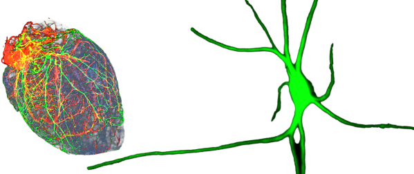
For the first time, we applied volume immuno-imaging combined with light sheet fluorescence microscopy to investigate a human brainstem nucleus called locus coeruleus, which is a key hub in the mammalian brain and exhibits perhaps the earliest tau pathology.
The study, which was published in Acta Neuropathologica, the leading journal of neuropathology, reveals an intriguing complexity and heterogeneity of tau cytoskeletal pathology in the human locus coeruleus already in very early stages of the Alzheimer’s disease spectrum. Moreover, it concludes that its special cytoarchitecture makes this tiny brain nucleus an ideal anatomical template for early accumulation and trans-neuronal spreading of pathological tau protein.
High resolution versions of supplementary videos of the Acta Neuropath paper are available here.
Further video demonstrations of human brain tissues from Alzheimer’s disease subjects are available here.
This paper received the ‘Publication of the Year award’ from the International Society of Alzheimer’s Research (ISTAART, Neuromodulatory Subcortical Systems Professional Interest Area) in 2023.
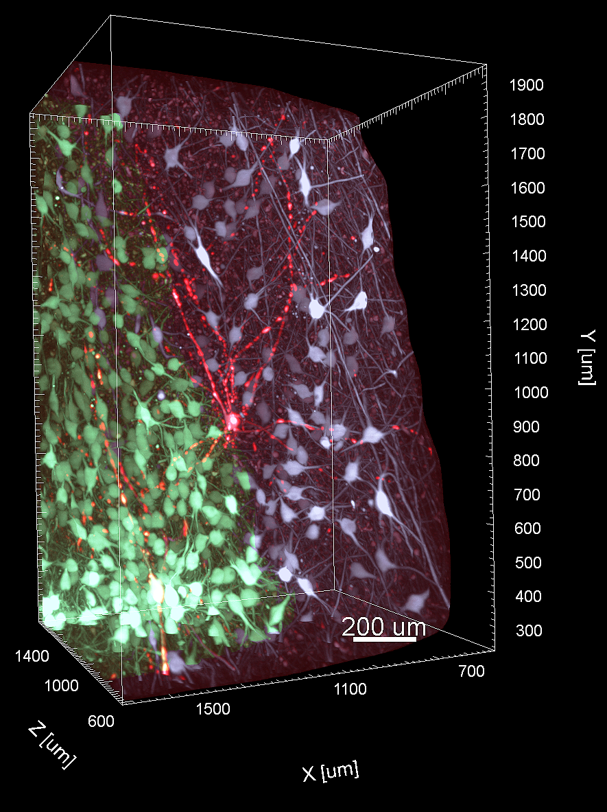
Hyperphosphorylated tau-bearing neuron (red) among tau-negative noradrenergic neurons (green and white cell populations) in the human locus coeruleus in 3D.