The neuropeptide galanin is necessary for the development of specific neuronal subsets. In this collaborative project, Dr. Joanna Baker and Dr. Zsofia Hevesi in the group of prof. Tibor Harkany (Medical University of Vienna and Karolinska Institutet Dept. Neuroscience) have explored the role of galanin in the peripheral‑to‑central connectivity in the somatosensory thalamus during whisker development.
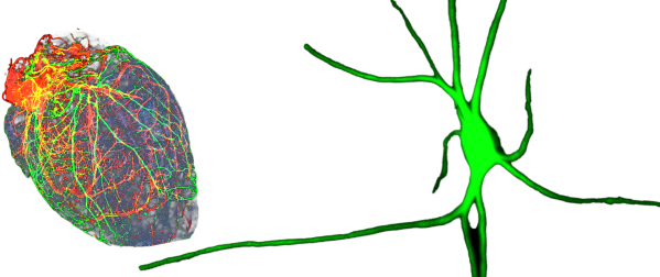
I contributed to this project with volume imaging of the mouse nucleus ventrobasalis thalami and its galanin-expressing neurons in various developmental stages, using anti-RFP volume immunostaining in gal-tdTomato+ mouse line.
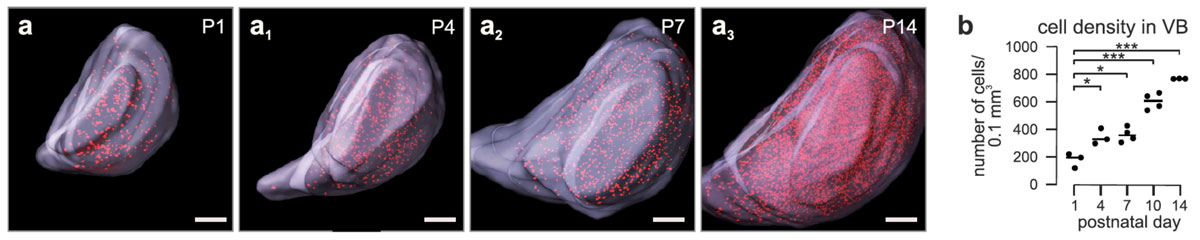
Volume imaging and galanin-expressing neurons in the developing mouse ventrobasal thalamus.
(a–a3 and b) Intact tissue imaging of the infant thalamus shows a progressive increase in cell density and the size of the nucleus.
RFP volume immunostaining, light sheet microscopy imaging, segmentation of n. ventrobasalis thalami based on tissue autofluorescence, 3D quantification of RFP+ cells, image production: Dr. Csaba Adori