Skip to content
Volume Imaging by Csaba AdoriVolume Imaging by Csaba Adori
  • Home
  • About me
  • iDISCO volume imaging
  • Scientific projects
  • Video demonstrations
  • Contact

HIGH RESOLUTION VERSIONS OF SUPPLEMENTARY VIDEOS IN THE PAPER OF GILVESY ET AL. (ACTA NEUROPATH, 2022).

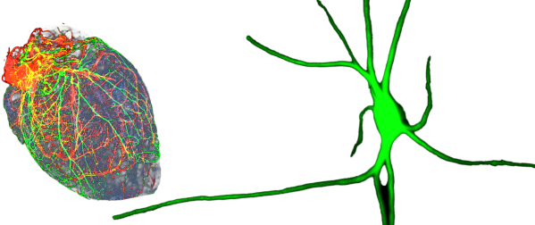
VIDEO #1: LC CORE FROM A BRAAK 0 BRAIN, TH VOLUME IMMUNOSTAINING

Description:

Segmented LC core, 2.42 μm x 2.42 μm x 2.50 μm voxel dimensions.

0:00 – 0:50 min.: Video demonstration of LC core cytoarchitecture.

0:50 – 1:10.: Demonstration of a small artery dorsally entering the LC core. The wall of the vessel is surrounded by densely packed TH+ neurites.

Download high. res. video

VIDEO #2: LC SHELL FROM A BRAAK 0 BRAIN, TH VOLUME IMMUNOSTAINING

Description:

300 mm x 900 mm x 2700 mm 3D crop, focusing to the LC shell. 1.51 μm x 1.51 μm x 2.00 μm voxel dimensions.

LC shell exhibits low TH+ cell density but a dense plexus of TH+ processes, mainly dendrites. Ventrally from the sparse shell TH+ neurons, long TH+ axons of the dorsal noradrenergic bundle are shown.

Download high. res. video

VIDEO #3: A4 FROM A BRAAK 0 BRAIN, TH VOLUME IMMUNOSTAINING

Description:

Segmented A4, 1.51 μm x 1.51 μm x 2.00 μm voxel dimensions.

A4 consisted of sparse TH+ cells with distinctly elongated somas. Their processes form a complex plexus around the mesenchephalic trigeminal tracts (me5). These processes are shown between 0:15 – 0:30. in details. Me5 is segmented and represented by a white surface between 0:00 – 0:07.

Download high. res. video

VIDEO #4: TAU CYTOSKELETAL PATHOLOGY IN THE LC OF A BRAAK NFT STAGE 0 BRAIN

Description:

Representative 3D crop from the LC core. AT8 channel. 2.42 μm x 2.42 μm x 2.50 μm voxel dimensions.

Sparse AT8+ cells with inhomogeneously or homogeneously filled somas and intact processes are dominating. AT8+ axon of a tau-bearing neuron is followed until the end of the 3D crop in between 0:47 – 1:06.

Download high. res. video

VIDEO #5: TAU CYTOSKELETAL PATHOLOGY IN THE LC OF A BRAAK NFT STAGE 1 BRAIN

Description:

Representative 3D crop from the LC core. AT8 channel. 1.51 μm x 1.51 μm x 2.00 μm voxel dimensions.

Strongly filled cells with partially atrophic dendritic tree are dominating. However, several transition forms are noticed in this sample.

Download high. res. video

VIDEO #6 TAU CYTOSKELETAL PATHOLOGY IN THE LC OF A BRAAK NFT STAGE 3 BRAIN

Description:

Representative 3D crop from the LC core. AT8 channel. 1.51 μm x 1.51 μm x 2.00 μm voxel dimensions.

Strongly filled cells with severely atrophic processes are dominating. Zoom to examples of  strongly filled cells with severely atrophic processes between 0:55 – 1:00.

Download high. res. video

VIDEO #7: TAU CYTOSKELETAL PATHOLOGY IN THE LC OF A BRAAK NFT STAGE 6 BRAIN (ALZHEIMER’S DISEASE)

Description:

Representative 3D crop from the LC core. AT8 channel. 1.51 μm x 1.51 μm x 2.00 μm voxel dimensions.

Abundant AT8+ mature tangle-like cells are dominating. Zoom to a neighboring tau-bearing cell duo between 0:38 – 0:43.

Download high. res. video

VIDEO #8: DORSO-VENTRAL DISTRIBUTION OF AT8+ VOLUME IMMUNOSTAINING IN THE LC

Description:

Segmented LC core from a Braak NFT stage 3 brain. TH (gray) and AT8 (red) channels. 4.83 μm x 4.83 μm x 4.00 μm (downsampled) voxel dimensions.

0:00 – 0:06 min.: TH and AT8 channels are shown.

0:07 – 0:40 min.: only AT8 channels is shown.

0:40 – 1:02 min.: Red and gray surfaces indicate AT8 volume staining in the dorsal and ventral halves of the LC core, respectively. Blue surface covers the entire LC core volume. Note the more abundant AT8 volume staining in the dorsal half.

Download high. res. video

VIDEO #9: DEMONSTRATION OF DENSE CELLS

Description:

3D crop from the LC core of a Braak NFT stage 2 brain. TH (gray) and AT8 (red) channels. 1.51 μm x 1.51 μm x 2.00 μm voxel dimensions.

0:00 – 0:05 min.: only TH channel, dense LC core cells are shown.

0:05 – 0:12 min: TH and AT8 channels are shown.

0:12 – 1:02 min.: only AT8 channel is shown; 0:40 – 0:45 zoom to a minigroup (formed by three cells with inhomogeneously filled soma and intact processes).  0:46 – 0:50: zoom to another minigroup, formed by (i) a cell with partially atrophic dendritic tree, (ii) a cell with severely atrophic processes and by (iii) cell with low intensity. 0:54 – 0:58: zoom to an AT8+ cell duo, formed by two cells with partially atrophic dendritic tree.

Download high. res. video

VIDEO #10: DENDRITES OF AT8+ LC NA CELLS EXTEND INTO THE SUBEPENDYMAL NA PLEXUS ALREADY IN BRAAK 0 STAGE

Description:

3D crop from a Braak NFT stage 0 brain block: dorsal-medial parts of the LC core, part of A4 as well as the periventricular area are shown. TH (gray) and AT8 (red) channels. 1.51 μm x 1.51 μm x 2.00 μm voxel dimensions.

Note the dense subependymal NA plexus below the 4th ventricle with sparse AT8+ processes.

0:00 – 0:06 min.: TH channel only.

0:06 – 1:10 min: TH and AT8 channels together.

Download high. res. video
Copyright 2025 © Volume Imaging / Dr. Csaba Adori

The website was designed by SOS Marketing

  • Home
  • About me
  • iDISCO volume imaging
  • Scientific projects
  • Video demonstrations
  • Contact